

Examinations
| Electrocardiogram (EKG) | Pulmonary function test | PET-CT | Cranial MRI scan |
| Abdominal ultrasound | Gastrocamera | Bone mineral density test | Cardiac ultrasound |
Electrocardiogram (EKG)
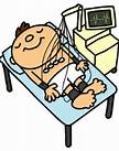
Resting electrocardiogram (EKG) Electrodes are attached to your wrists, ankles, and chest, and the electric signals from the heart are recorded. Electricity is not sent through the body, so there is no paralysis or pain. The examination is performed while lying on a bed and resting, and is finished in a few minutes. Arrhythmia, cardiac hypertrophy, cardiac infarctions, and other heart diseases can be discovered with this examination. |
Pulmonary function test
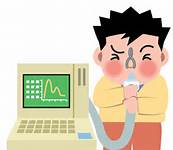
Lung capacity, forced expiratory volume in one second, forced expiratory volume one second percent
By breathing in and out through a mouthpiece, we can examine your lung functions. There is no pain, but you must be able to make your best effort during the examination. The capacity of your lungs and forced vital capacity are measured, and diseases of the lungs and air duct can be found through this examination. |
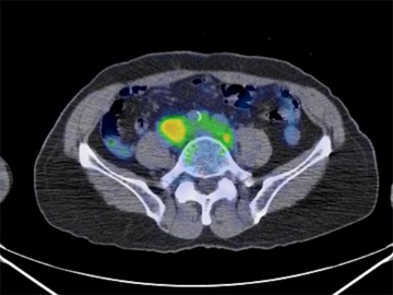
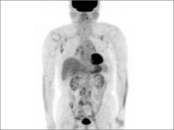
PET-CT
|
PET stands for Positron Emission Tomography. A medicine is injected into a vein in your arm to expel the positrons (protons) in your body, and the way the medicine collects in parts of your body is seen through a special camera. CT scans are done in the same way. PET and CT scans typically scan the area from around your ears all the way to your ankles. PET/CT machines create 2 images at roughly the same time, and a high-quality image that is valuable for diagnosis can be created through combined both images.
For our PET/CT scans, we inject fluorodeoxyglucose (FDG), a medicine that labels fluorine-18 (18 F) radioactive isotopes, which collects in places such as cancer cells that consume large amounts of glucose, which allows us to see if cancer is present or not. Cancerous cells require 3 to 8 times the glucose compared to normal cells. The medicine also collects in pneumonia and abscesses (an area filled with pus), so a collection of medicine does not necessarily mean cancer.
|
Cranial MRI

・Cranial MRI & MRA and Cervical MRA ・Consultation (Neurological Assessment) and Explanation of Results
Strokes, cerebral hemorrhages, and subarachnoidal hemorrhages can end lives in an instant, and it is not uncommon to suffer serious side effects such as speech disorders and paralysis, as well. It is important to take preventative measures as it is too late once symptoms have appeared. We offer a Cranial MR Checkup to make early discoveries of any potential threats.
Diseases that can be discovered early on ・Stroke with no subjective symptoms ・Unruptured cerebral aneurysms ・Brain tumors ・Cerebral A‐V malformations ・Cerebral hemorrhages ・Moyamoya disease, etc. |
Abdominal ultrasound
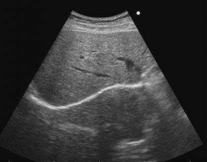
Upper endoscopy (ultrasound examination: liver, bile duct, renal gland, spleen, and pancreas) (An upper endoscopy examines the liver, bile duct, renal gland, spleen, and pancreas. If food remains in the stomach, this causes bowel movements and gas buildup, leading to an ineffective examination. This examination requires fasting at breakfast on the day of the examination. A lubricant jelly is applied to the stomach, and moving the device around, the liver, bile duct, renal gland, spleen, and pancreas are examined, checking for cancer, polyps, or any other lesions. Obesity can sometimes prevent the pancreas from being examined.)
|
Gastrocamera
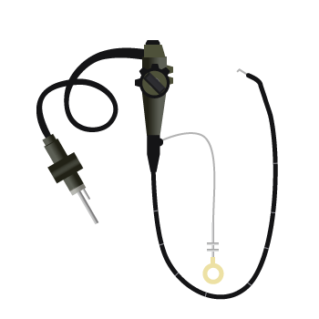
A camera is inserted from the mouth for a general examination or to examine the cause of symptoms such as abdominal pain, heartburn, and anemia. The condition of the stomach and duodenum is displayed directly on the monitor, allowing ulcers, inflammation, tumors, and polyps to be discovered. During the examination, a tissue sample from the affected area may be taken to be examined later (microscopic examination). The endoscope has been greatly reduced in size thanks to advances in technology, leading to a much less painful examination. We also offer a nasal endoscope (inserted through the nose) examination that uses a very small endoscope, reducing the vomiting reflex during the examination. |